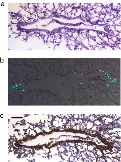
Fig. 1.
AA deposition in foie gras. (a) Large venule surrounded by residual, extensively vacuolated fatty hepatic tissue (hematoxylin/eosin stain). (b) Green birefringent amyloid deposits in the blood vessel wall (Congo red stain). (c) Immunohistochemical identification of vascular AA amyloid. (Scale bar, 62 μm.)

