Figure 4.
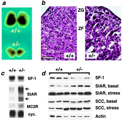
Altered histology and gene expression in SF-1 +/− adrenal cortex. (a) Adult SF-1 +/+ adrenals are significantly larger than +/− adrenals (total adrenal weight for males: +/+, 3.3 ± 0.06 mg; +/−, 1.4 ± 0.02 mg; P < 0.01, n = 8 per group; females: +/+, 6.5 ± 0.06 mg; +/−, 2.3 ± 0.08 mg; P < 0.01, n = 7 per group). Body weight is equivalent in SF-1 +/+ and +/− mice (data not shown). Adrenal cross-sectional area is 2–3 times smaller in SF-1 +/− embryos compared with +/+ embryos at both E15.5 (+/+, 0.148 ± 0.001 mm2; +/−, 0.043 ± 0.001 mm2; P ≤ 0.01) and E18.5 (+/+, 0.216 ± 0.004 mm2; +/−, 0.090 ± 0.001 mm2; P ≤ 0.01). (b) Histological analysis of SF-1 +/+ and +/− adrenals. SF-1 +/− adrenals display cortical cell hypertrophy (cells per 0.01 mm2: +/+, 90.1 ± 0.9; +/−, 66.1 ± 0.7; P ≤ 0.01), dilated adrenocortical sinusoids, and a hypoplastic zona fasciculata (ZF) adjacent to the zona glomerulosa (ZG). Bar = 50 μM. (c) Northern blot analyses of SF-1, StAR, and MC2-R mRNA expression in SF-1 +/+ and +/− adrenals. Whereas SF-1 +/− adrenals express lower levels of SF-1 mRNA, they express higher levels of both the 3.4- and 1.6-kb StAR transcripts and the 1.7-kb MC2-R transcript. Cyclophilin mRNA levels (cyc.) are shown for loading comparison. (d) Western blot analyses of SF-1, StAR, SCC, and actin protein expression in adrenals from basal (fed) or stressed (fasted) SF-1 +/+ and +/− mice.

