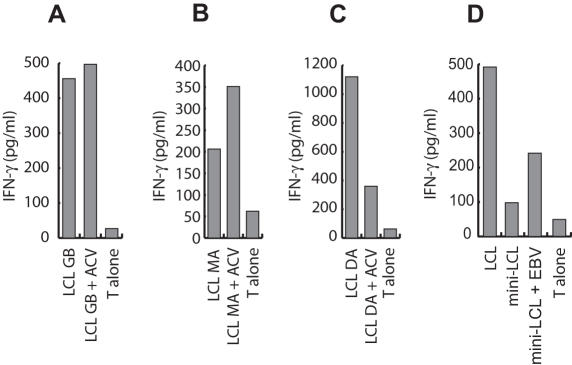
Figure 6. Presentation of virion antigens is impaired in acyclovir-treated LCL.
LCL either left untreated or treated with acyclovir for two weeks were used as targets for BMRF1 (A), autoantigen (B), or BNRF1-specific T cells (C). Acyclovir treatment neither affected presentation of the autoantigen nor the EBV early lytic cycle antigen BMRF1, but severely reduced the presentation of the virion antigen BNRF1. (D) T cell lines generated by repeated stimulation of peripheral blood CD4+ cells with acyclovir-treated LCL recognized LCL and mini-LCL that had been pulsed with purified EBV particles, suggesting that late lytic cycle antigen-specific T cells still expand under these stimulation conditions.

