
Fig. 4.
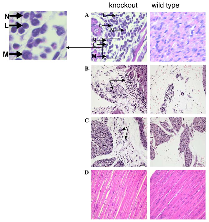
Pathological analysis of neonatal mice with and without trsp in muscle. Left panels show tissues and organ from affected pups (designated knockout) and right panels, the corresponding tissues and organ from unaffected pups (designated wild type). Tissue analyzed, ages of neonatal mice, and the fold magnifications were: (A) heart, 10 days (40×); and letters with arrows designate: L, lymphocyte; N, neutrophil; M, macrophage; (B) mediastinum, 6 days (20×); (C) mediastinum, 10 days (20×) and (D) skeletal muscle, 10 days (40×). Panel descriptions are given in the text. Staining was carried out as given in Section 2.