
Figure 6.
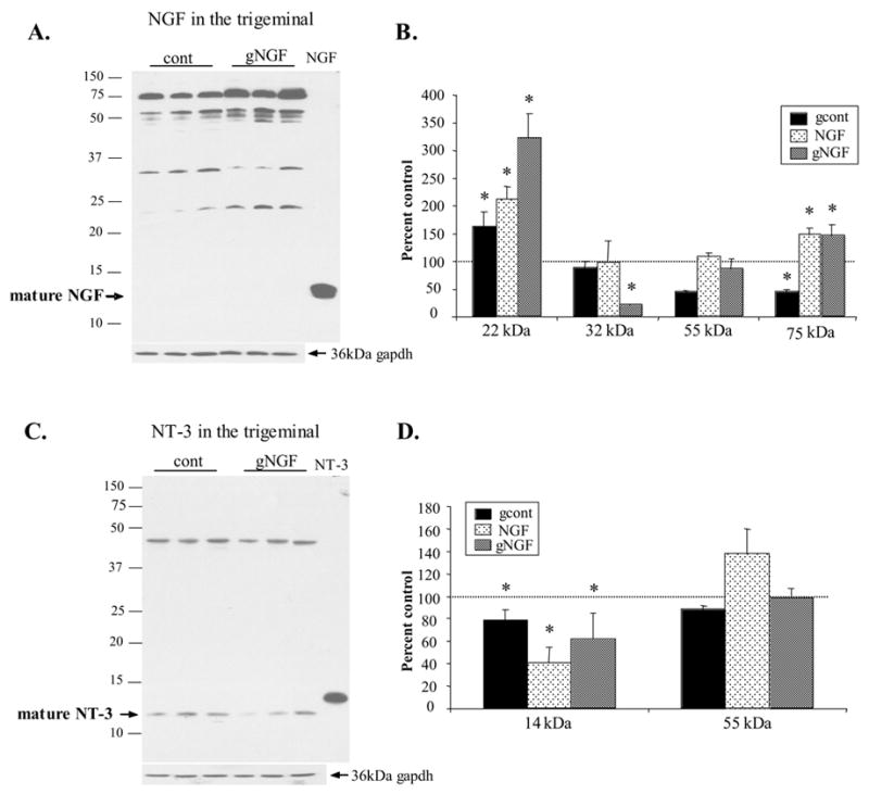
NGF and NT-3 species in the trigeminal ganglion following sympathetic denervation (gcont), 2 week NGF administration (NGF), and denervation followed by 2 week NGF administration (gNGF). A. Representative NGF western blot of trigeminal ganglion from gNGF animals (gNGF) and control animals (cont) revealed the presence of the 22, 32, 55, and 75kDa NGF proteins. B. Semi-quantitative analysis revealed a significant increase in the 22kDa NGF species in all three treatment groups. The 75kDa species was decreased following denervation but NGF administration resulted in an increase in this species. 20μg protein loaded per lane. Each lane represents a different animal. C. Representative blot reveals the presence of the mature NT-3 and a 50 kDa NT-3 species. The mature NT-3 form was decreased when sympathetic denervation was followed by NGF administration (gNGF) when compared with controls. B. Semi-quantitative analysis reveals a significant decrease in ‘mature’ NT-3 in all three treatment groups, suggesting that the reduction of NT-3 at the targets was not a result of increased transport to the trigeminal ganglion. The 50 kDa species was decreased in gcont treatment only. 20μg protein loaded per lane. Each lane represents a different animal. (*p<0.05 compared with control)