
Figure 6.
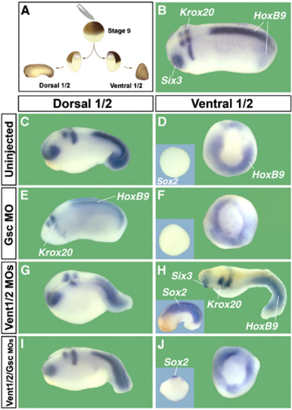
Knockdown of Gsc and Vent1/2 restores normal development of dorsal and ventral half-embryos (n=52 or more per experimental set). (A) Embryos were bisected into dorsal and ventral halves at blastula stage. (B) Control sibling at the same magnification as the other panels. (C, D) Bisectioned control embryos form smaller but well-proportioned dorsal half-embryos, whereas ventral halves differentiate into belly-pieces that express HoxB9 in the ventral mesoderm (Wright et al, 1990) but are devoid of neural tissue, as indicated by the lack of Sox2 expression (inset). (E, F) Gsc depletion (136 ng MO) causes a reduction of the head in dorsal halves, whereas ventral halves are not affected. (G, H) Dorsal halves of Vent1- and Vent2-depleted embryos (45 ng each) are dorsalized, but retain overall DV patterning. The corresponding ventral halves are strongly dorsalized, including expression of spinal cord (HoxB9), brain (Krox20, Six3), and pan-neural Sox2 marker (inset). (I, J) Remarkably, both halves of triple knockdown embryos (45 ng each) develop as the uninjected control half-embryos.