
Figure 3.
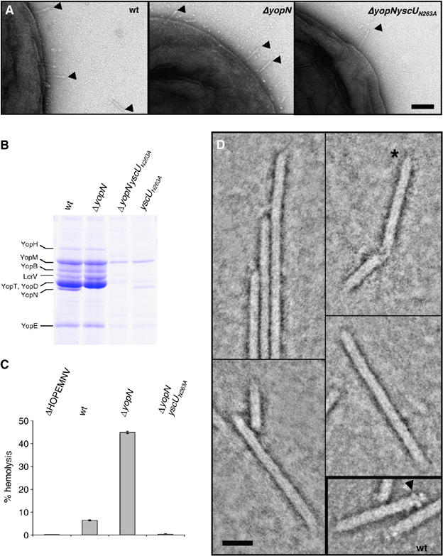
The yscUN263A mutant does not induce hemolysis in RBC due to a missing tip complex. (A) Transmission electron micrographs of Y. enterocolitica E40 WT, ΔyopN and ΔyopNyscUN263A bacteria negatively stained with 2% uranyl acetate. Scale bar, 100 nm. Needles are indicated by arrowheads. (B) Yops secreted by Y. enterocolitica wt, ΔyopN, yscUN263A and ΔyopNyscUN263A bacteria. Coomassie-stained 12% SDS–PAGE. The position of YopN is indicated on the left. (C) Percentage lysis of RBCs after 1 h of contact with the indicated Y. enterocolitica strains. (D) STEM dark-field image of Y. enterocolitica yscUN263A needles. Protein is displayed in bright shades. Inset: wt needles similarly imaged. The yscUN263A mutant needles had rather pointed ends (asterisk); the tip complexes so characteristic of wt needles (arrowhead) were not detected. Scale bar, 20 nm. Strains: wt (pYV40); ΔyopN (pIM41); ΔyopNyscUN263A (pISO4010); yscUN263A (pISO4007); ΔHOPEMNV (pMN4002).