
Figure 5.
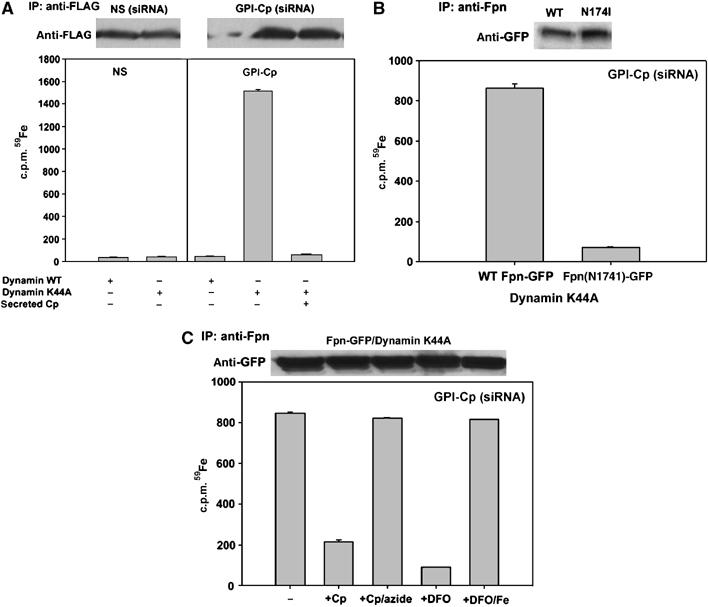
Iron is bound to Fpn in the absence of GPI-Cp. (A) Cells transfected with nonspecific (NS) or GPI-Cp-specific oligonucleotides were then transfected with Fpn-FLAG and dynamin-GFP or dynamin(K44A)-GFP. Twenty-four hours after transfection (72 h post siRNA), cells were incubated for 12 h with Tf (59Fe)2±2.0 μM Cp, washed and Fpn-FLAG was immunoprecipitated using anti-FLAG resin. Fpn-FLAG was eluted with 0.2 mg/ml FLAG peptide, the immunoprecipitates assessed by Western blot and the amount of Fpn bound 59Fe determined. (B) C6 glioma cells silenced for GPI-Cp were transfected with Fpn-GFP or Fpn(174)-GFP and dynamin(K44A)-GFP. Twenty-four hours post-transfection (72 h post GPI-Cp siRNA), cells were treated as in (A), samples immunoprecipitated with rabbit anti-Fpn antibody and protein A/G resin and the amount of immunoprecipitated 59Fe-Fpn determined. (C) Cell lysates from Fpn-GFP/dynamin(K44A)-GFP expressing cells were incubated with Cp, azide-treated Cp, DFO or iron-loaded DFO for 6 h at 4°C. Fpn was immunoprecipitated as in (B) and the amount of 59Fe associated with Fpn-GFP was determined.