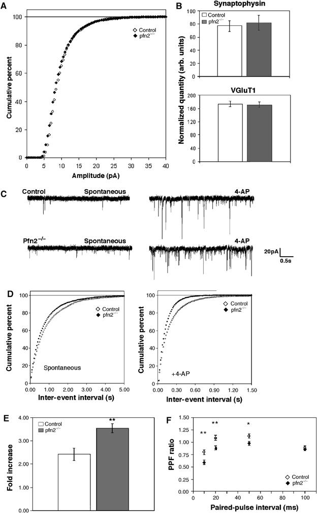
Figure 6.

Glutamatergic neurons in pfn2−/− mice are hyper-excitable and show higher vesicle release probability. (A) Cumulative percentage curves of the amplitude distribution of miniature EPSCs in pfn2−/− (n=7/3: number of cells/number of mice) and control neurons (n=5/3). (B) Quantification of synaptophysin and VGluT1 levels (synaptic content) in striata by Western blot (n=6 for each genotype). (C) Representative traces from a control and a pfn2−/− neuron before and after application of 4-AP illustrate the hyper-excitability of the mutant neurons. (D) Cumulative percentage curves of the average inter-event interval distribution of spontaneous EPSCs in basal conditions (spontaneous) and after application of 4-AP (+4-AP, control n=8/3, pfn2−/− n=7/2; Kolmogorov–Smirnov test P=0.037 and P<0.0001, respectively). (E) Excitability of pfn2−/− neurons in response to calcium increase. Evoked vesicle release upon change of [Ca2+]/[Mg2+] ratio from 1 to 4 increased the response amplitude 3.5-fold in pfn2−/− neurons, while in control neurons the increase was 2.4-fold (P=0.0055 in Student's t-test; n=5/3 for control and n=5/4 for pfn2−/− mice). (F) PPF in control and pfn2−/− littermates. Pfn2−/− neurons showed stronger depression that correlates with a higher release probability at presynaptic sites (n=10/5 for control and n=11/4 for pfn2−/− mice). *P<0.05, **P<0.01, ***P<0.001. Error bars represent s.e.m.
