
Figure 7.
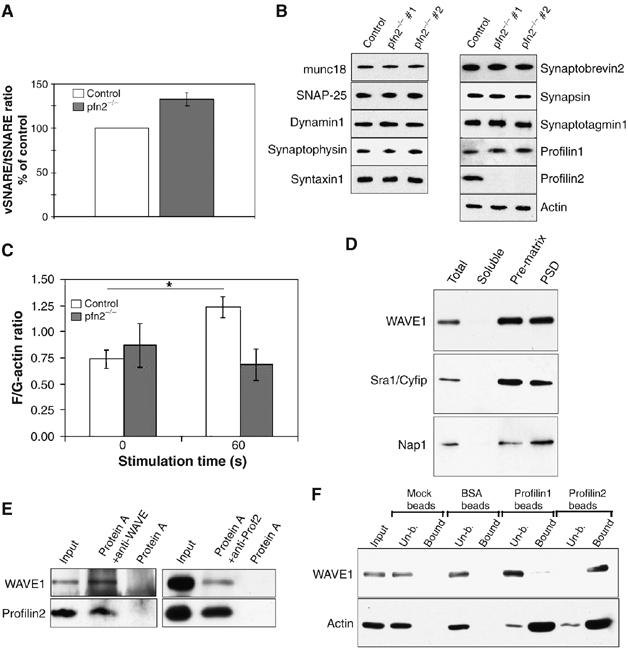
Lack of actin polymerization in pfn2−/− synaptosomes correlates with increased vesicle docking/priming. Specific binding of the WAVE-complex to profilin2. (A) Synaptic vesicle priming based on vSNARE/tSNARE ratios after immunoprecipitation of tSNARE (syntaxin1) from pfn2−/− synaptosomes is increased by 30%. vSNARE/tSNARE ratios from control mice (n=5) were set to 100% and the mutant ratios (n=4) expressed accordingly. (B) Synaptosomes from control and pfn2−/− mice were analyzed by Western blot using a panel of antibodies detecting different markers of the vesicle release machinery. Two different mutant mice are shown to account for sample variability. (C) Synaptic F/G-actin ratios were not significantly different in resting synaptosomes (0 s), after 60 s stimulation with 20 mM K+ the F-actin/G-actin ratio raised in the control, while no significant increase was seen in the mutants (n=3 for mutant and control; ANOVA, interaction genotype x KCl stimulation: F[1,8]=5.365, P=0.049). (D) Distribution of WAVE-complex components in synaptosomal fractions. WAVE1, Sra1/Cyfip, and Nap1 were found enriched in the presynaptic matrix and the PSD. (E) In synaptosomal lysate, profilin2 co-immunoprecipitated with WAVE1 (left panel) and WAVE1 co-immunoprecipitated with profilin2 (right panel). (F) Specific binding of WAVE1-complex to profilin2. Pull down of WAVE1 from cortical protein extracts using profilin1- and profilin2-coupled beads shows specificity for profilin2.