
Figure 4.
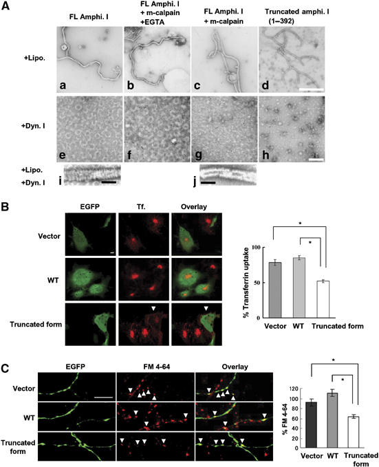
Amphiphysin I cleaved by calpain and a truncated form (1–392) are capable of tubulation with liposomes, but do not form ring structures with dynamin I (A: a–j), and inhibitory effect of calpain-dependent truncated form of amphiphysin I on transferrin uptake in Cos-7 cells (B) and FM 4–64 labeling in primary culture of hippocampal neurons (C). (A: a–d) The effect of truncation on the ability of amphiphysin I to form tubules with liposomes. As a chelator of Ca2+, EGTA inhibits calpain activity. (A: e–h) The effect of both forms of amphiphysin I on the formation of ring structures with dynamin I. (A: i, j) Comparison of helical collars on the tubules of FL and truncated amphiphysin I when incubated with dynamin I plus liposomes. Bars in A: d and h, 500 nm; bars in A: i and j, 100 nm. (B) Confocal laser scanning micrographs of: left panel, EGFP expression on COS-7 cells transfected with pIRES2-EGFP vector only (Vector), the vector containing wild-type amphiphysin I cDNA (WT) and the vector containing truncated amphiphysin I (1–392) cDNA (truncated form); middle panel, transferrin (Tf.) uptake in similarly transfected cells exposed to transferrin 24 h later; and, right panel, showing the overlay of EGFP and transferrin uptake. Transferrin uptake was inhibited in cells transfected with truncated amphiphysin I (arrows); bar, 10 μm. For quantification of the results, transferrin uptake was calculated as the mean fluorescence density for 315–660 cells under each condition and the mean values (±s.e.m.) were expressed as a percentage of normal transferrin uptake in untransfected cells. *P<0.01. (C) Fluorescence imaging micrographs of: left panel, EGFP expression on presynapses of neurons transfected with vector, WT amphiphysin I and the truncated form (amphi. 1–392); middle panel, subtracted images of FM 4–64 (loading minus unloading); right panel showing overlay of EGFP expression and FM 4–64 labeling. Bar, 10 μm. FM 4–64 labeling was inhibited in boutons transfected with truncated amphiphysin I (arrows). Right graph shows the results of quantitative analysis. FM 4–64 labeling was calculated as the integral fluorescence intensity for 47–55 boutons under each condition and the mean values (±s.e.m.) were expressed as a percentage of normal FM 4–64 labeling in untransfected boutons. *P<0.01.