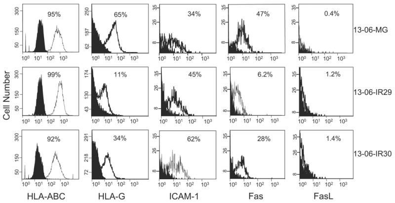
FIGURE 4.

Expression of classic HLA-ABC class I antigen, nonclassic HLA-G, ICAM-1, and Fas and FasL by parental 13-06-MG glioma cells and 13-06-IR29 and 13-06-IR30 IR clones by flow cytometric analyses. The histograms show the nonspecific binding of isotype and secondary antibodies (filled peaks) and the degree of specific staining (unfilled peaks). The percentages of positive cells are indicated and the relative antigen densities can be ascertained by placement and height of the peak on the abscissa.
