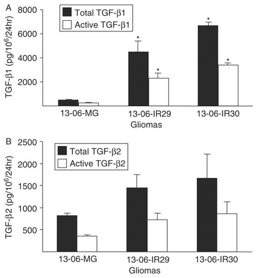
FIGURE 7.

Detection of TGF-β isoforms secreted by monolayers of the parental glioma cells or IR clones. The rates of total (latent plus active forms) and active forms of (A) TGF-β1 and (B) TGF-β2 secretions were determined by ELISA. Rates of TGF-β secretion are expressed as pg TGF-β/106 cells/24 h ± SEM. Asterisks indicate statistically significant production (P ≤ 0.02) relative to the parental glioma cells by the nonparametric Kruskal-Wallis test.
