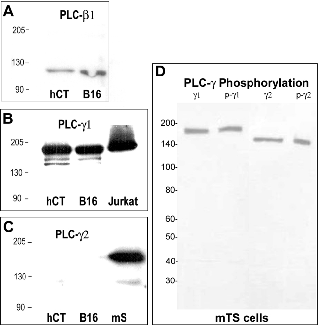
Figure 3.
Specificity of antibodies against PLC isoforms. Western blotting was conducted, as described in the Materials & Methods, using antibodies against PLC-β1 (panel A), PLC-γ1 (panel B) or PLC-γ2 (panel C). The lanes contained proteins extracted from human HTR-8/SVneo cytotrophoblast cells (hCT), mouse B16 melanoma cells (B16), mouse spleen (mS; positive control for PLC-γ2) and a commercial Jurkat cell lysate preparation (Jurkat; BD Biosciences, San Jose, CA; positive control for PLC-γ1). In panel D, antibodies against PLCγ1 (γ1) and PLCγ2 (γ2) were compared to antibodies recognizing the tyrosine phosphorylated forms of each enzyme (p-γ1, p-γ2) using a lysate prepared from mouse TS cells. Molecular weights (kDa) are indicated to the left in each panel.

