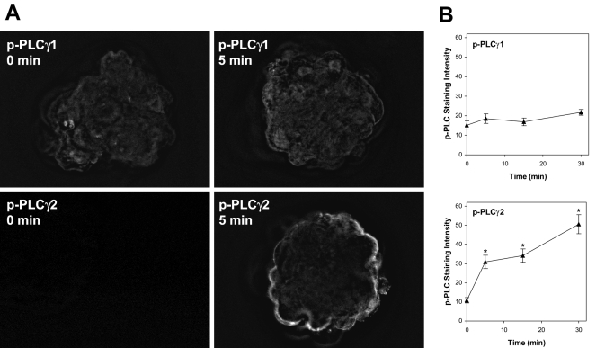
Figure 7.
Phosphorylation of PLC-γ1 and PLC-γ2 during exposure to FN-120. Adhesion-competent blastocysts cultured to GD7 were exposed to FN-120 for 0, 5, 15 or 30 min and then fixed, permeablized, and labeled by immunofluorescence using an antibody against either PLC-γ1 phosphorylated at tyrosine 783 or PLC-γ2 phosphorylated at tyrosine 1217. Panel A shows deconvolved images of embryos exposed to FN-120 for 0 or 5 min. Panel B shows the specific phospho-PLC staining quantified as described in the Materials and Methods section for each antibody over a 30 min period of exposure to FN-120. The mean ± SEM is shown at each time for 10-12 embryos. *, P < 0.05, compared to Control.

