
Figure 2.
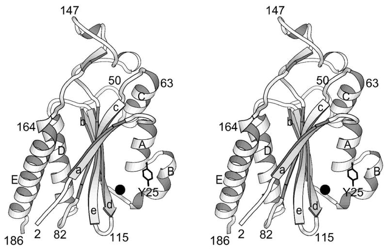
Ribbon drawing of minMobA. This stereo picture illustrates the fold of the protein and displays its secondary structural elements. Residues along the backbone are labeled to aid in following the polypeptide path. The active site Mn atom and Tyr side chain are also shown.