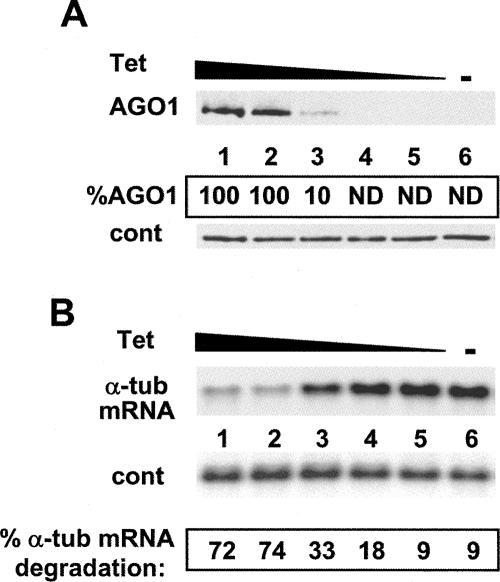
FIGURE 2.
(A) Disappearance of AGO1 protein in response to decreasing tet concentrations. TbAGO1KOc cells were grown in the absence of tet for 4 d, and then incubated for 24 h with different tet concentrations (lanes 1–5) or without tet (lane 6). Cytoplasmic extracts were analyzed by Western blotting with an anti-AGO1 antiserum. Tet concentrations: (lane 1) 10 μg/mL; (lane 2) 1.0 μg/mL; (lane 3) 0.1 μg/mL; (lane 4) 0.01 μg/mL; (lane 5) 0.001 μg/mL. (Cont) A cross-reactive protein. The amount of AGO1 was quantitated from the autoradiogram and is expressed as the percent of AGO1 expressed at the highest tet concentration. (ND) Not detectable. (B) The RNAi competency of TbAGO1KOc cells decreased in response to incubation with decreasing amounts of tet. TbAGO1KOc cells were grown using the same tet concentrations as described in A. After 24 h, the cells were electroporated with 2 μg of α-tubulin dsRNA and then returned to the medium for 2 h. Ten micrograms of total RNA was analyzed by Northern blotting using an α-tubulin coding-region probe. The hybridized membranes were analyzed by PhosphorImaging, and the percent α-tubulin mRNA degradation was calculated, after adjusting for loading differences, by taking the amount of α-tubulin mRNA present at time 0 as 100%.

