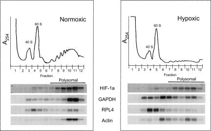
FIGURE 3.
Polysome distribution of the HIF-1α, GAPDH, RPL4, and the actin mRNAs are differentially affected by hypoxic treatment. PC-3 cells were grown under normoxic and hypoxic conditions (1.0% O2 20 h) and subjected to polysome analysis. Polysome lysates were fractionated by sucrose gradient centrifugation and collected into twelve 1 mL fractions. Absorbance profiles (A254) are shown at the top of each panel. The top of the gradient is on the left and peaks representing the 40 S and 60 S ribosomal subunits are denoted. The nonpolysomal region of the gradient included fractions 1–6 and the polysomal portion is fractions 7–12. RNA was isolated from each fraction and subjected to Northern Analysis using 32P-labeled probes to detect the HIF-1α, GAPDH, RPL4, and actin mRNAs.

