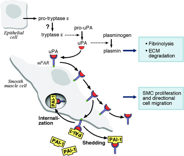
Figure 7.
Schematic model of the tryptase ε–mediated activation of uPA/uPAR-dependent reactions. In this model, active tryptase ε converts pro-uPA to uPA (red symbol) in the epithelium. uPA converts plasminogen to plasmin which, in turn, promotes fibrinolysis and the degradation of extracellular matrices. Some uPA binds to the uPAR (blue symbol) on the surface of the smooth muscle cell and induces varied signaling events in the cell. The uPA/uPAR-activated cells eventually increase their expression of the inhibitory factor PAI-1 (yellow symbol), and the exocytosed serpin binds to the uPA/uPAR complex to dampen the protease-mediated activation response. Some of the multimeric complex is internalized and endocytosed uPA is rapidly destroyed. The remainder is shed in a phospholipase C–dependent manner from the plasma membrane with or without PAI-1. In some cells, uPA can directly bind to the αMβ2.48 Thus, the uPA that is generated by tryptase ε also could regulate certain integrin-dependent signaling events. ECM indicates extracellular matrix.

