Figure 2.
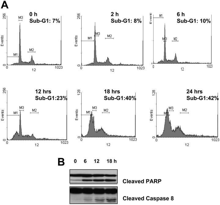
Seliciclib treatment induces apoptosis of MM cells in a time- and dose-dependent manner. Cell-cycle analysis by PI staining was performed on MM1.S cells cultured with media alone or seliciclib (25 μM) for the specified time points. Seliciclib resulted in an increase in sub-G1 fraction as early as 12 hours, with 42% of the cells in sub-G1 phase at 24 hours (A). This was associated with an increase in PARP and caspase 8 cleavage (B). M1 indicates sub-G1 gate; M2, G2M gate; and M3, G1 gate.

