Figure 4.

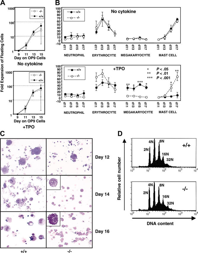
Myeloid-lineage development of B-raf–/– ES cells cocultured with OP9 cells. (A) B-raf–/– (–/–) and wild-type (+/+) hematopoietic cells from day 9 of OP9 coculture were replated on a fresh OP9 layer, and the number of viable floating and loosely adherent cells generated on days 11 to 15 in the absence (top) or presence (bottom) of 50 ng/mL TPO was determined using trypan blue dye exclusion. The fold expansion for each day is plotted on log scale. (B) Cell differentials of the floating cells generated on days 11 to 17 in the absence (top) or presence (bottom) of TPO (50 ng/mL) were determined by microscopic examination of Giemsa-stained cells. The y-axis indicates the percentage of each lineage cell type. Immature blastic cells and macrophages were also detected in floating cells on day 11 and days 13 to 17, respectively (data not shown). (A-B) The data represent the average ± SD for 3 independent experiments. (C) Cell morphology of wild-type and B-raf–/– ES-derived hematopoietic cells developed on days 12 to 16 of coculture in the presence of 50 ng/mL TPO is presented using cytospun cells stained with Diff-Quick. Original magnification, × 200;× 1000 for insets, which highlight the mast cells. Images were obtained using a Leica DMLB microscope (Leica, Bannockburn, IL) and ScionImage software (ScionImage, Frederick, MD), and were cropped with Adobe Photoshop. (D) DNA ploidy distribution for B-raf–/– (–/–) and wild-type (+/+) ES-derived CD41+ cells shows no significant difference between the 2 genotypes, with DNA ploidy ranging from 2N to 32N for each. One of 3 representative DNA ploidy experiments is shown.
