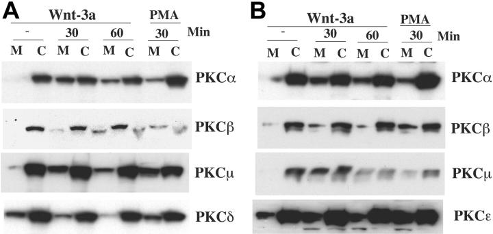
Figure 5.
Wnt-3a alters cellular localization of PKCs α, β, and μ. H929 (A) and OPM-2 (B) cells starved in serum-free medium for 12 hours were treated with Wnt-3a or con-CM for the indicated time. Cell lysates from cytosolic (C) or membrane (M) fraction were prepared as described in “Material and methods.” Lysates were resolved on 8% SDS-PAGE gels, transferred to membranes, and blotted with indicated antibodies. Treatment of cells with 100 nM PMA (phorbol 12-myristate 13-acetate) for 30 minutes was used as a positive control for PKC membrane translocation.

