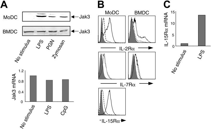
Figure 1.
Regulation of γc cytokine receptors and Jak3 in DCs. (A) Jak3 expression in activated DCs. Human MoDCs (top) and murine BMDCs (middle and bottom) were stimulated with LPS (1 μg/mL), PGN (5 μg/mL), or zymosan A (5 μg/mL) for 24 hours. Cell lysates (100 μg) were electrophoresed and blotted with anti-Jak3 antibody. Jak3 mRNA was assessed by real-time PCR in murine BMDCs stimulated with LPS (1 μg/mL) or CpG oligonucleotides (oligos) (1 μg/mL) for 6 hours (bottom). (B) Human MoDCs and murine BMDCs were stimulated with LPS (1 μg/mL) for 24 hours, stained with FITC-labeled antibodies, and then analyzed by flow cytometry. Broken line indicates unstimulated cells; solid line, activated cells. Cells stained with an isotype control antibody are depicted in the shaded histogram. (C) IL-15Rα expression by murine BMDCs was measured by real-time PCR in cells treated with LPS (1 μg/mL) for 6 hours or left unstimulated.

