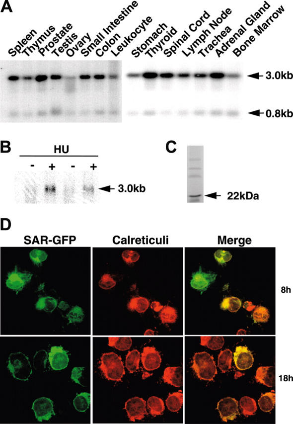
Figure 1.

SAR mRNA expression in different tissues and HU induction of SAR. (A) mRNA blots of multiple human tissues were hybridized with a 32P-labeled SAR-specific probe. (B) Human adult erythroid cells were treated with (+) or without (-) 100 μM HU, as detailed in “Materials and methods.” Total RNA was isolated on day 10 and probed for SAR mRNA by Northern blotting. (C) In vitro transcription/translation of the SAR protein produced a 22-kDa peptide. (D) Cellular localization of SAR tagged with GFP at the 3′ end in SAR-transfected K562 cells. Green indicates GFP fluorescence; red indicates staining for calreticulin, which indicates the endoplasmic reticulum; yellow (in the merged image) represents overlapping green and red fluorescence. Cells were incubated for 8 or 18 hours.
