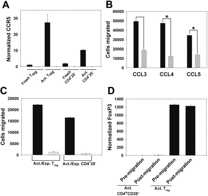
Figure 1.
Functional expression of CCR5 on Tregs. (A) QPCR assessment of CCR5 expression in column-purified T CD25- T cells from WT (▪) and CCR5-/- ( ) mice. Shown is CCR5 expression in freshly isolated cells, and cells cultured as in “Materials and methods.” Data are mean ± the standard error of the mean (SEM). (B) WT Tregs (▪) and CD4+CD25- T cells (
 ) were cultured as in panel A, and migration in response to 10 ng/mL CCL3, CCL4, and CCL5 was determined. Data are mean ± SEM. *P < .05. (C) Chemotaxis of WT (▪) and CCR5-/- (
 ) Tregs and CD4+CD25- T cells, cultured as above, in response to 100 ng/mL CCL4. Data are mean ± SEM. (D) QPCR analysis of FoxP3 mRNA expression in cultured WT Tregs and CD4+ regs and CD4+ CD25- T cells prior to chemotaxis and in cells that migrated in response to 100 ng/mL CCL4. Data are mean ± SEM.

