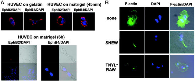
Figure 4.
Surface EphB2 and EphB4 expression and actin polymerization during cord formation. (A) HUVECs were plated onto gelatin-coated or Matrigel-coated glass chamber slides and incubated at 37°C. After 45 minutes and 6 hours of incubation, cells were fixed with 1% formaldehyde, stained for EphB2 or EphB4 with specific goat IgG antibodies followed by Texas Red donkey anti–goat IgG and DAPI, and examined with a confocal system. Images were from phase-contrast and epifluorescence microscopy showing representative HUVECs cultured on gelatin (18 hours) or Matrigel (45 minutes and 6 hours) (original magnification, ×10 ×25). (B) HUVECs were plated onto Matrigel-coated glass chamber slides and were incubated at 37°C in medium alone, medium with the SNEW peptide (100 μM), or medium with the TNYL-RAW peptide (100 μM) for 6 hours and then stained with phalloidin-FITC and DAPI. Images from confocal epifluorescence microscopy show representative F-actin detection in HUVECs (original magnification, ×25).

