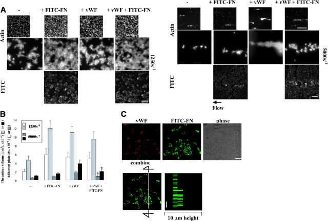
Figure 6.
Effect of perfused VWF and fibronectin on thrombogenesis on collagen under shear conditions and localization of VWF and fibronectin. A suspension of platelets and red blood cells with or without 50 μg/mL FITC-fibronectin (FITC-FN) and 200 μg/mL unlabeled fibronectin and/or 10 μg/mL VWF was perfused over surfaces coated with 20 μg/mL collagen at a wall shear rate of 1250 s–1 or 5000 s–1 for 5 minutes. (A) Coverslips were taken out of the chamber, stained with rhodamine-phalloidin, and processed for epifluorescence microscopy. The small and large pictures were taken with 10 × (bar = 100 μm) and 100 × (bar = 10 μm) objectives, respectively. (B) Thrombus volume and number of adherent platelets were calculated as described in Figure 3. Values represent the mean ± SD (n = 3-4). *P < .05 compared with when the perfusate contained just VWF. (C) After perfusion, nonpermeabilized platelets were incubated with mouse anti-VWF followed by rhodamine-conjugated anti–mouse IgG antibody. Confocal microscopy was performed as described in “Materials and methods.” The images represent a 1-μm slice taken from 4-μm height of the coverslip. The bottom right panel shows a perpendicular section through the line. Bar = 10 μm.

