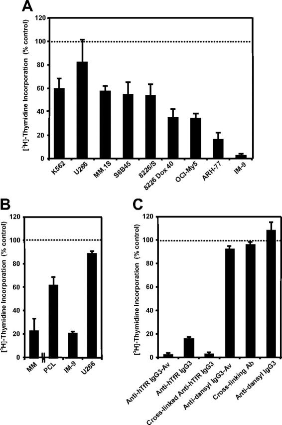
Figure 1.

Anti-hTfR IgG3-Av inhibits the growth of malignant hematopoietic cells. (A) Proliferation assay of a panel of hematopoietic malignant cell lines incubated with 34 nM anti-hTfR IgG3-Av for 72 hours. Cells were incubated with 4 μCi (0.148 MBq)/mL [3H]-thymidine for an additional 24 hours prior to harvesting. Data represent the mean of quadruplicate samples of 2 independent determinations of [3H]-thymidine incorporation. (B) Proliferation assay of primary cells isolated from the bone marrow of a patient with MM and a patient with PCL. Myeloma cells from the patients were isolated in 2 independent experiments by positive selection. As a control, 2 cell lines, IM-9 (highly sensitive) and U266 (less sensitive), were tested in parallel. Control data from the experiment with the PCL patient are shown. All cells were treated in triplicate with 100 nM anti-hTfR IgG3-Av for 48 hours and then incubated with 4 μCi (0.148 MBq)/mL [3H]-thymidine for an additional 48 hours prior to harvesting and determination of radioactivity. (C) ARH-77 cells were incubated with 11 nM anti-hTfR IgG3-Av, anti-hTfR IgG3, anti-hTfR IgG3 cross-linked with a 5-fold excess of secondary Ab, secondary Ab alone, the anti-dansyl IgG3 isotype control, or the anti-dansyl IgG3-Av for 72 hours. Cells were incubated with 4 μCi (0.148 MBq)/mL [3H]-thymidine for an additional 24 hours prior to harvesting and determination of radioactivity. Data shown are representative of 3 independent experiments. All data are presented as the percentage [3H]-thymidine incorporation compared with control cells treated with buffer. Error bars indicate the standard deviation.
