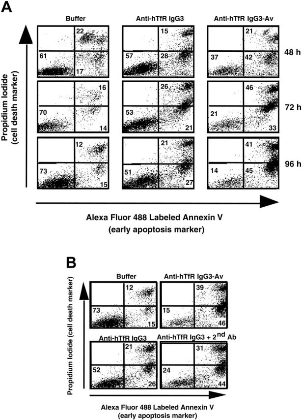
Figure 2.

Anti-hTfR IgG3-Av induces apoptosis in ARH-77 cells. (A) Cells were incubated with 11 nM anti-hTfR IgG3 or anti-hTfR IgG3-Av for the indicated times. Cells were then washed, stained with annexin V-Alexa Fluor 488 and PI, and analyzed by flow cytometry. Data shown are representative of 3 independent experiments. (B) ARH-77 cells were treated with 1.2 nM anti-hTfR IgG3-Av, 3.7 nM anti-hTfR IgG3, or 3.7 nM anti-hTfR IgG3 cross-linked with a 5-fold excess of secondary Ab for 96 hours and analyzed by flow cytometry. The percentage of cells located in each quadrant is shown in the corner. Results are representative of 2 independent experiments.
