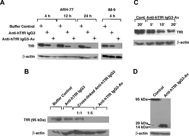
Figure 4.
Anti-hTfR IgG3-Av induces the highest levels of intracellular degradation of the TfR. (A) ARH-77 and IM-9 cells were treated with buffer, 11 nM anti-hTfR IgG3, or 11 nM anti-hTfR IgG3-Av. ARH-77 was treated for 4, 12, and 24 hours, while IM-9 was treated for 4 hours. (B) ARH-77 cells were treated with buffer, 11 nM anti-hTfR IgG3, 11 nM anti-hTfR IgG3 cross-linked with secondary mAb in 1:1 or 1:5 ratios, or 11 nM anti-hTfR IgG3-Av for 8 hours. (C) ARH-77 cells were treated with buffer or 11 nM anti-hTfR IgG3-Av for 5, 10, and 20 minutes. (D) ARH-77 cells were treated with buffer or 11 nM anti-hTfR IgG3-Av for 4 hours. In all cases, cells were harvested, and whole-cell lysates were prepared and analyzed by immunoblotting with a murine mAb against the intracellular N-terminus of the human TfR followed by incubation with goat anti-mouse IgG-HRP conjugate. The blot was then stripped and reblotted with a murine anti-β-actin mAb. In panels A, B, and C, only the regions of the 95-kDa intact TfR and β-actin are shown. In panel D, the blot from 14 to 95 kDa is shown. These experiments were repeated at least twice with similar results.

