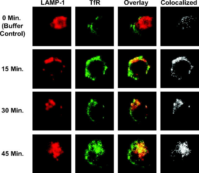
Figure 6.
Anti-hTfR IgG3-Av directs the TfR to an intracellular compartment containing the lysosomal marker LAMP-1. ARH-77 cells treated with 11 nM anti-hTfR IgG3-Av for 0 (buffer control), 15, 30, or 45 minutes were fixed, permeabilized, and stained as described in “Materials and methods.” Cells were then analyzed by confocal microscopy. The image of a representative cell from each time point is shown immunostained for TfR (green) and LAMP-1 (red) with overlay. The area where colocalization occurs is shown in white. The percent colocalization was calculated as described in “Confocal microscopy” (Table 1).

