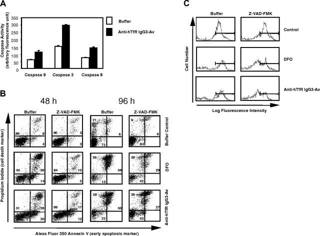
Figure 8.
Anti-hTfR IgG3-Av induces caspase activation, mitochondrial depolarization, and a partially caspase-independent cell death in ARH-77 cells. (A) Cells treated with buffer or 50 nM anti-hTfR IgG3-Av for 48 hours were lysed and caspase activities measured by specific fluorogenic substrates. Each value is the mean of quadruplicate samples. (B) Cells preincubated with or without 100 μM Z-VAD-FMK were treated with buffer, 50 μM DFO, or 50 nM anti-hTfR IgG3-Av for 48 or 96 hours. Cells were then washed and stained with DiOC6(3), annexin V-Alexa Fluor 350, and PI, and analyzed by flow cytometry. The annexin V/PI profiles of treated cells at 48 and 96 hours are shown with the percentages of cells located in each quadrant. (C) Nonapoptotic cells (lower left quadrants of panel B) at 48 hours were gated and their mitochondrial membrane potentials shown in log fluorescence intensity. These experiments were repeated in both ARH-77 and IM-9 cells with similar results.

