Figure 3.
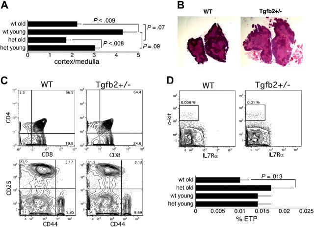
Analysis of the involuting thymus in Tgfb2+/- and wt mice. (A) Ratio of cortical to medullary surface area in Tgfb2+/- (het) and wt mice (n = 12 sections from 3 thymi; young are 8 weeks old, old are 14-16 months old). (B) Representative example of hematoxylin and eosin-stained thymi from 16-month-old wt and Tgfb2+/- mice (original magnification, × 10). (C) Representative example of staining of thymi from wt and Tgfb2+/- mice for CD4 and CD8 (top panels) and for CD25 and CD44 (bottom panels, gated on cells negative for CD3, CD4, CD8α, CD8β, B220, Ter119, NK1.1, Gr-1, Mac1). (D) Representative example of the detection of ETPs (lin- CD25-c-kit+IL7Rα-/lo, plots gated on cells that were negative for CD25, CD3, CD8α, CD8β, TCRαβ, TCRγδ, NK1.1, CD19, Mac1, GR1, B220 and Ter119) in wt and Tgfb2+/- thymi (top panels), and frequency of ETPs in thymi from young (2-6 months) and old (14-16 months) Tgfb2+/- mice and wt littermates (n = 4 litters in young mice and 5 litters in old mice; 2 mice from each genotype and each litter were pooled for each analysis).

