
Figure 3.
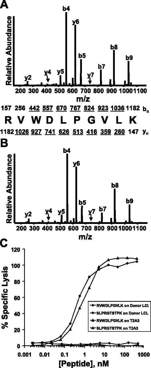
Identification of the antigenic peptide recognized by the CTL clone CTL-7A7. (A) CAD mass spectrum of candidate peptide (M+2H)2+ ion with monoisotopic m/z of 591.865 as eluted from HLA-A*0301-homozygous mHAg+ EBV-LCLs. (B) CAD mass spectrum of synthetic peptide RVWDLPGVLK. Mass spectra were recorded on a Finnigan LCQ ion trap MS operating with a 3.0 atomic mass unit isolation window and 35% collision energy. The b and y ions are labeled above and below the amino acid sequence, respectively. Ions observed in the spectrum are underlined. (C) CTL-7A7 mHAg epitope reconstitution with synthetic RVWDLPGVLK peptide. 51Cr-labeled donor EBV-LCLs or T2A3 target cells were pulsed for thirty minutes at 37°C with the indicated concentrations of synthetic peptides RVWDLPGVLK or SLPRGTSTPK, then tested for recognition by CTL-7A7 CTLs in a 4-hour 51Cr-release assay at an E/T ratio of 10:1. The control peptide SLPRGTSTPK corresponds to the HLA-A*0301-restricted epitope recognized by CTL clone DRN-7 (E.H.W., manuscript in preparation).