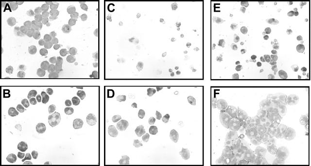
Figure 6.
Morphology of 32DA1 cells in different growth conditions. 32DV4 (A,C,E) and 32DA1.3 cells (B,D,F) were grown in IL-3 (A-B), without cytokine (C-D), or in G-CSF (E-F). Photomicrographs of cells attached to glass slides using a Cytospin apparatus and then stained with Wright/Giemsa stain are shown.

