Figure 6.

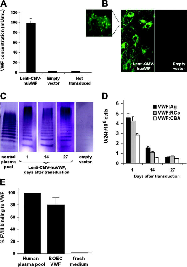
Characterization of huVWF expression by canine type 3 VWD BOECs transduced with Lenti-CMV-huVWF. (A) HuVWF levels in conditioned medium from transduced VWD BOECs as determined by ELISA (1 day after transduction) or from negative controls (ie, type 3 VWD BOECs transduced with empty vector or not transduced). (B) HuVWF immunostaining in cytoplasm and Weibel-Palade bodies (magnification) of VWD BOECs transduced with Lenti-CMV-huVWF or empty vector. (C) Multimer analysis was performed on 130 ng VWF present in normal human plasma pool and on expression medium harvested on 1, 14, and 27 days after transduction from canine VWD BOECs transduced with Lenti-CMV-huVWF or empty vector. (D) At several time points after transduction, 24-hour conditioned media from transduced VWD BOECs were tested for VWF:Ag levels by the VWF:Ag ELISA and for functional VWF using the VWF:RCo ELISA and the VWF:CBA assay. No VWF activity was found in conditioned medium from VWD BOECs transduced with empty vector. (E) Capacity of VWF expressed by transduced VWD BOECs to bind FVIII as determined by ELISA. Fresh medium was used as a negative control. Data are mean ± SEM; n = 3.
