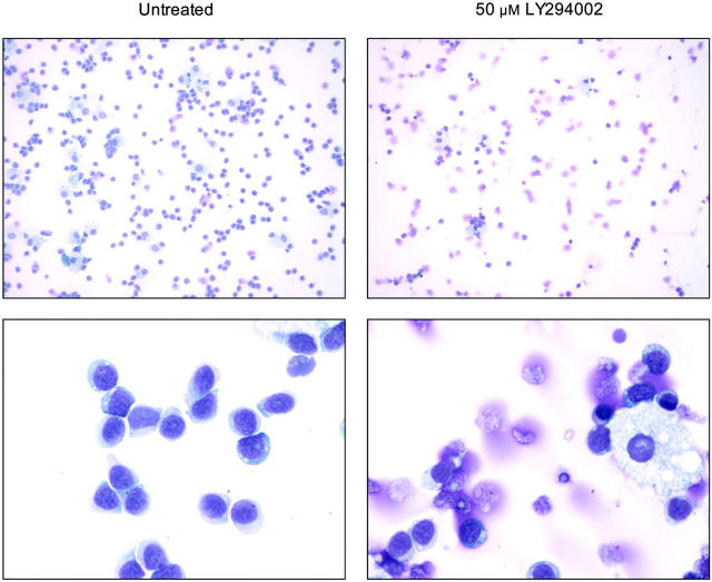
Figure 5.
PI3K inhibition induces morphologic changes in T-LGL consistent with apoptosis. Untreated (control) T-LGL are noted in the left panels intermixed with occasional macrophages. The right panels show images of Wright-stained T-LGL PBMCs that had been treated for 20 hours with LY294002 (50 μM). Apoptotic cells are seen in the LY-treated T-LGL. Original magnifications: low power, × 200; high power, × 1000 (oil).

