Figure 6.
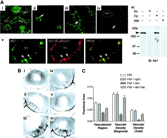
Ab1 labels CD148 on murine endothelium and inhibits corneal angiogenesis in mice. (Ai-v) Mouse cryostat tissue sections were immunostained with FITC-conjugated Ab1. Ab1 labels arterial and capillary endothelium (arrows) of various tissues, including kidney (i), cerebellar cortex (ii), endocardium (iii), and corneal angiogenic vessels (iv) following stimulation with a basic FGF–impregnated hydron pellet (ppt). Highly magnified image of renal artery exhibits endothelial localization of CD148, which overlaps VE-cadherin (arrowheads, v). Art indicates renal artery; glom, renal glomerulus; and peri, peritubular capillary. Images were taken using a Zeiss LSM 410 laser-scanning microscope (Carl Zeiss) with Zeiss 10×/0.3 numerical aperture (NA), 40×/1.3 NA, and 63×/1.3 NA objective lenses, and with Image Browser 5 LSM ver3.2 (Carl Zeiss). Figure panels were prepared with Adobe Photoshop 7.0 (Adobe Systems). Original magnifications: 100× (i), 400× (ii), 400× (iii), 400× (iv), and 1200× (v). (vi) Mouse kidney tissue lysates (300 μg) were subjected to immunoblotting using HRP-conjugated Ab1 preabsorbed with CD148 recombinant proteins (50 μg) (CD148ec indicates ectodomain; CD148cy, cytoplasmic domain) or epitope peptide (10 μg) (pep). (B) Hydron pellets impregnated with (i) vehicle (PBS), (ii) bFGF (3.0 pM), (iii) bFGF + control mouse IgG1 (74 pM), (iv-v) bFGF + Ab1 (74 pM), or (vi) bFGF + Ab1 Fab fragment (74 pM) were implanted and photographed at 5 days after implantation. (C) The graph displays means ± SEM (n = 8, excluding IgG1 controls [n = 6]) for the fractional areas vascularized, the vascular density within that region, and the vessel density as a fraction of the total corneal image area, as indicated in the figure.

