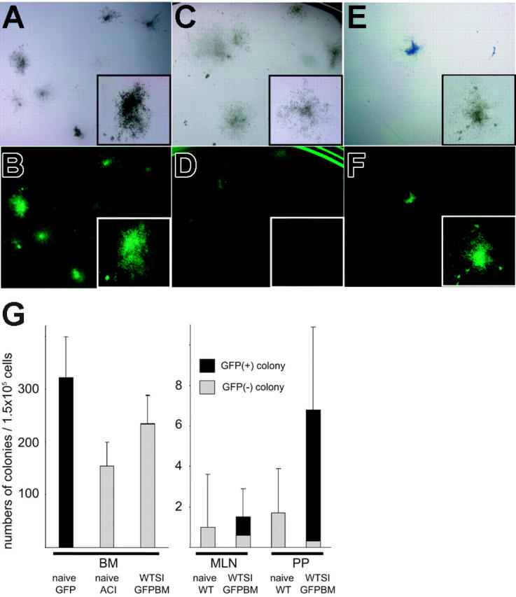
Figure 6.

Colony-forming unit in culture (CFU-C) assay of isolated cells from WT intestine grafts cotransplanted with GFP-positive BM. (A-B) Naive GFP-positive BM formed more than 300 colonies per 1.5 × 10⁵ cells, and all of them were GFP positive under fluorescent microscopy. (C-D) BM cells taken from the recipient of WT intestine plus GFP-positive BM formed comparable numbers of colonies as normal ACI animals, but very few GFP-positive colonies were found despite stable macrochimerism. (E-F) Cells isolated from PPs and MLNs of WT intestine grafts transplanted with GFP-positive BM developed GFP-positive colonies with a frequency of 2 to 6 per 1.5 × 10⁵ cells. CFU-Cs of cells isolated from naive WT intestinal PPs and MLNs were 1 to 3 per 1.5 × 10⁵ cells. (G) Mean numbers of colonies per 1.5 × 10⁵ cells (n = 3). Panels A, C, and E are under an inverted microscope; B, D, and F are under an Olympus SZ×12 fluorescence dissecting microscope with magnification wheel set at 7×, insets at 30×. SI indicates small intestine.
