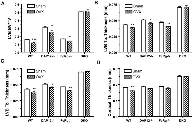
Figure 2. OVX-induced bone remodeling in lumbar vertebra and cortical bone.
Six weeks after operations, the sixth lumbar vertebra (LVB6), and tibia were processed and analyzed by µCT, as described in Materials and Methods. Trabecular bone volume/tissue volume (A), trabecular thickness (B), and trabecular number (C) of LVB6, and cortical thickness of tibia at distal tibiofibular junction (D) are shown. WT: wild-type mice; DKO: mice lacking both DAP12 and FcRγ. N = 5 in each group. *: p<0.05; **: p<0.01; ***: p<0.001; when compared to SHAM groups, respectively.

