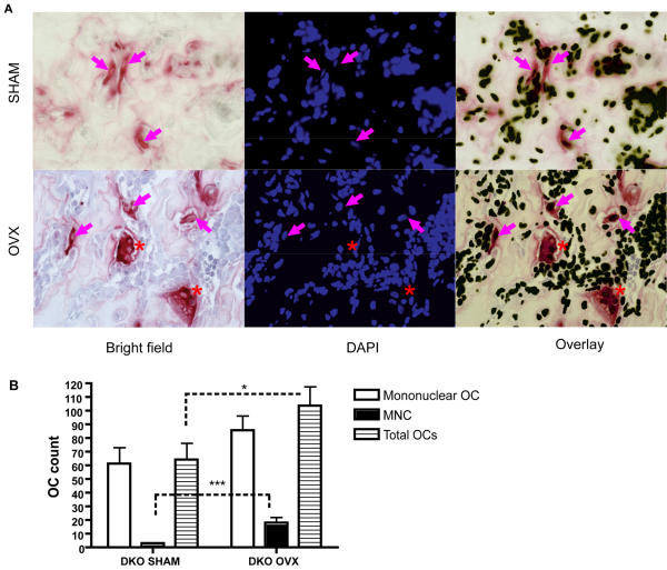
Figure 4. Increased osteoclast formation in vivo in DAP12-/-FcRγ-/- OVX group.
(A) TRACP staining and DAPI staining was performed on paraffin section of decalcified femur of SHAM and OVX groups of DKO mice. Brightfield and fluorescence images were taken with 40× objective. Red staining in brightfield indicates TRACP+cells. Magenta arrow indicates mononuclear TRACP+ osteoclasts; red asterisk indicates TRACP+ multinuclear osteoclasts (MNCs). (B) Mononuclear, multinuclear, and total osteoclast number per section was counted. *: p<0.05; ***: p<0.001; when compared between groups indicated by dotted line. N = 3 each group.

