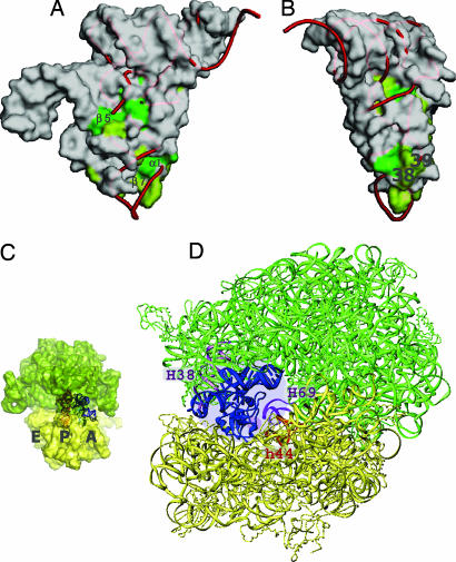
Fig. 4.
Functional mimicry of a canonical tRNA by tmRNA and SmpB. (A and B) Molecular surface of SmpB, represented as with tmRNA. The conserved residues on the surface of SmpB are colored green (complete) or light green (partial). The tRNAPhe was superimposed on the tRNA domain of the tmRNA by using the nucleotides of the acceptor stems and the T arms, and the central cores with the residues of SmpB corresponding to the nucleotides (Fig. 2C). Positions 38 and 39 of the tRNA are labeled in B. (C and D) tmRNA with SmpB on the ribosome. The 50S and 30S subunits are colored green and yellow, respectively. tmRNA and SmpB are superimposed on the A-site tRNA of the 70S ribosome (16). The yeast tRNAPhe is shown in the P-site of the ribosome in C. The corresponding region of SmpB for nucleotide positions 38 and 39, shown in A and B, is indicated by a small purple circle around H69 and h44 of the ribosome in D.

