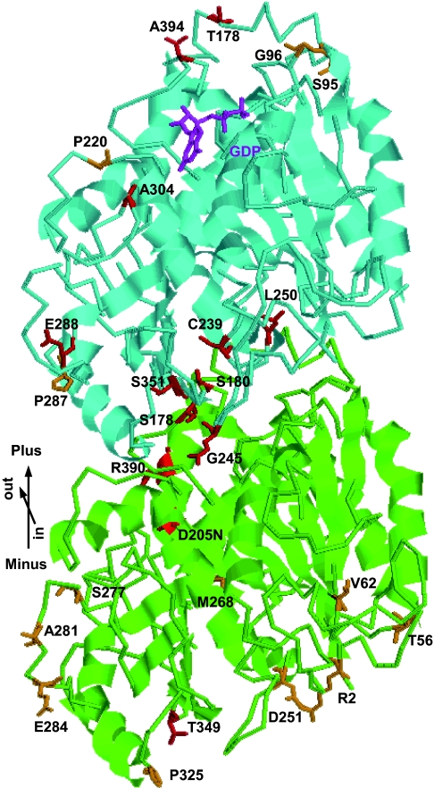
Fig. 2.
Tubulin mutations generally map to the interacting regions. Tubulin heterodimers are viewed from the microtubule lumen side and with their β-subunits facing the plus end. Residues mutated in the right-handed helical growth mutants are shown in orange, whereas those in left-handed mutants are in red. α-tubulin, green; β-tubulin, cyan; and GDP bound in β-tubulin, magenta. The model is based on a structural model of pig tubulins (14, 15). Note that S95 in β-tubulin corresponds to S96 in TUB1 due to a single amino acid insertion in the N terminus of TUB1.

