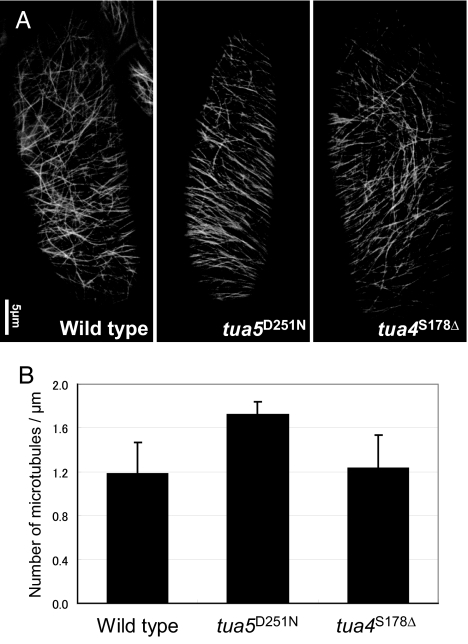
Fig. 4.
Microtubules in tua5D251N hypocotyl cells are more abundant and align more transversely. (A) Cortical microtubules in epidermal cells of 4-day-old hypocotyls, visualized by GFP-TUB6. (B) Number of microtubules in unit length. Cortical microtubules that crossed the mid-line of a cell's long axis were counted.

