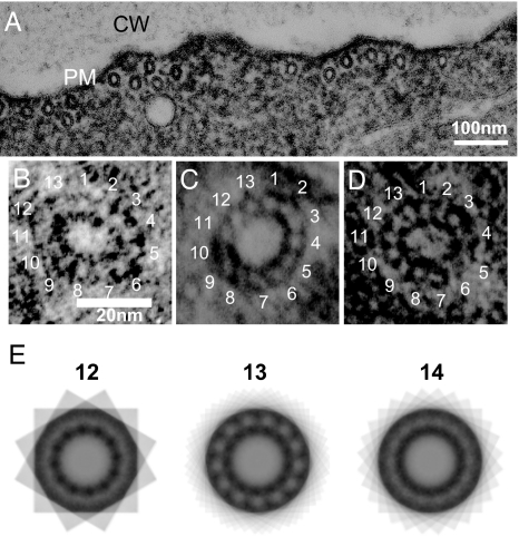
Fig. 6.
Transverse section of cortical microtubules. (A) Electron micrograph of a longitudinal section of a root epidermal cell in the elongation zone. Cortical microtubules are seen associated with the plasma membrane (PM). CW, cell wall. (B–D) Transverse sections of cortical microtubules from the wild type (B), tua5D251N (C), and tua4S178Δ (D). (E) Multiple exposures of a tua5D251N microtubule, where n = 12, 13, or 14 equal arcs of a complete circle were used. The strongest reinforcement of the ordered pattern occurred when n = 13.

