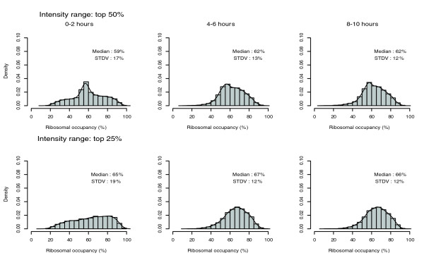
Figure 5.

Analysis of ribosomal occupancy. Histograms of ribosomal occupancy, which is defined as the percentage of polysomal associated mRNAs for individual transcript species. The top panels include the probe sets with the top 50% signal intensity among all the probe sets and the bottom panels only include the probe sets with the top 25% intensity. The x-axis is the value of ribosomal occupancy (%). The spacing of rectangles is 5%. The heights of rectangles (y-axis) are densities, defined as density = (number of genes with their value within the 5% range of individual rectangle/total number of genes in the set)/width (5% in these plots). Therefore, the total area of all rectangles equals 1. The lines over the rectangles are the smoothed histograms. STDV, standard deviation.
