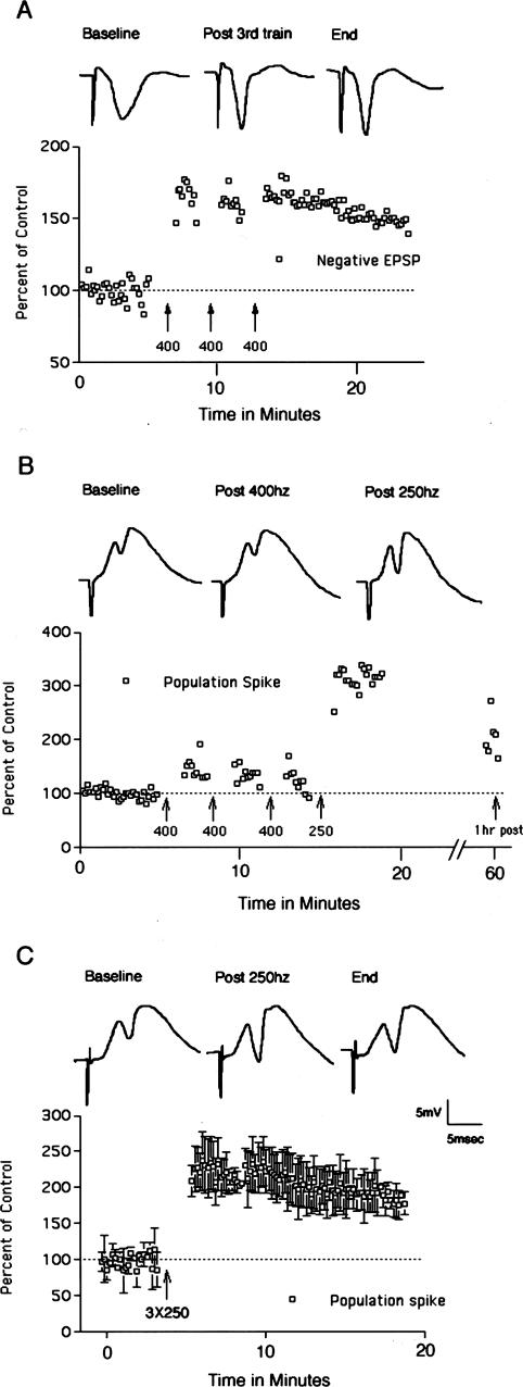
Figure 3.
Perforant path LTP in mice. (A) Perforant path LTP induced by 400 Hz stimulation. Traces illustrate negative-going population EPSPs recorded by a micropipette positioned in the molecular layer of the dorsal blade of the dentate gyrus. The graph illustrates EPSP amplitude as a percent of baseline before and after 400 Hz stimulation. (B) Perforant path LTP is induced more readily by 250 Hz than 400 Hz stimulation. In this experiment, three bouts of 400 Hz stimulation did not induce LTP, whereas three bouts of 250 Hz stimulation at the same intensity was effective in inducing LTP. Traces illustrate positive-going population EPSPs and population spikes recorded by a micropipette positioned in the cell body layer of the dorsal blade of the dentate gyrus. The graph illustrates population-spike amplitude as a percent of baseline before and after 400 Hz and then 250 Hz stimulation. Data points on the far right indicate population-spike amplitude 1 h after inducing LTP with 250 Hz stimulation. (C) Average LTP induced by 250 Hz stimulation in three experiments. The graph illustrates the average population-spike amplitude as a percent of baseline ± SD. Traces illustrate positive-going population EPSPs and population spikes recorded by a micropipette positioned in the cell body layer of the dorsal blade of the dentate gyrus in a representative animal.

