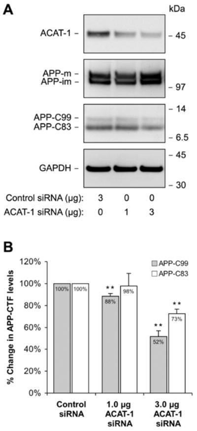
Figure 2. Decreased ACAT-1 Expression Levels Correlate with Reduced Proteolytic Processing of APP.

H4APP751 cells were transfected with either control siRNA (mouse ACAT-1) or siRNA specific for human ACAT-1 and analyzed 96 h later for ACAT-1 expression and APP proteolytic fragements (A). The levels of the APP C-terminal fragments (APP-C99 generated by β-secretase and APP-C83 produced by α-secretase) were quantitated in four independent experiments (B). The values were normalized to GAPDH and the immature form of APP holoprotein (APP-im). * p < 0.05, ** p < 0.01.
