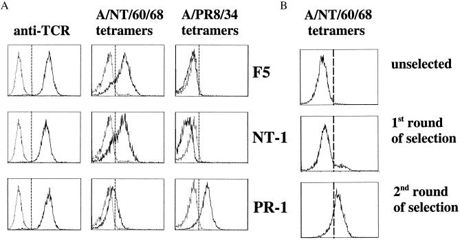
Figure 2.
MHC tetramer analysis of in vitro selected TCRs. (A) Flow-cytometric analysis of 34.1Lζ cells expressing the F5 (Top), NT-1 (Middle), or PR-1 TCRs (Bottom). The left panels represent staining with phycoerythrin-conjugated anti-TCRβ chain (H57–597) mAb. The middle panels represent staining with APC-labeled tetrameric H-2Db complexes containing the A/NT/60/68 NP epitope (ASNENMDAM), and the right panels represent staining with APC-labeled H-2Db tetramers containing the A/PR8/34 NP epitope (ASNENMETM). Tetramer staining was performed at 37°C (37). (B) Selection of influenza A-reactive TCRs from in vitro TCR libraries. The panels represent staining of the TCRβ CDR3 library with APC-labeled tetrameric H-2Db complexes containing the A/NT/60/68 NP epitope before screening (Top) and after 1 (Middle) and 2 (Bottom) sorts with A/NT/60/68 H-2Db tetramers. Tetramer selections were performed at 4°C.

