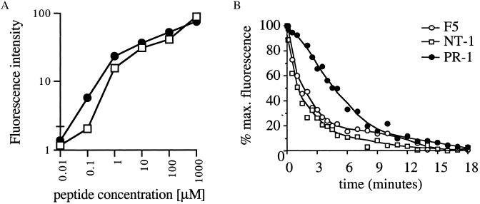
Figure 4.
TCR-MHC off-rates of in vitro selected TCRs. (A) Comparison of H-2Db-binding affinity of the A/NT/60/68 (▫) and A/PR8/34 (●) peptide. TAP-deficient RMA-S cells (39) were incubated overnight at 37°C with peptides at the indicated concentrations. Cells were stained with biotin-conjugated anti-mouse H-2Db antibodies (PharMingen) and phycoerythrin-conjugated streptavidin and analyzed by flow cytometry. Fluorescence intensity is calculated as: FIexp − FI0. Data shown are means of triplicates ± SD. (B) Determination of MHC-TCR dissociation rates. 34.1Lζ TCR-expressing cells were stained with their cognate APC-labeled peptide/H-2Db tetramers at 4°C and subsequently exposed to an excess of homologous unlabeled H-2Db monomers at 25°C. Decay of H-2Db-tetramer staining was measured by flow cytometry and is plotted as the percentage of maximum staining.

